
Why "my back hurts" is a poor starting point
One painful region can have ten different origins. Pain behind the knee can come from the meniscus, from short hamstrings, from referred lumbar pain, or from a Baker's cyst. Each takes a different path, and an exercise programme built for one can be useless — or harmful — for another.
What standard clinical tests do is narrow the possibilities. No single test is conclusive, but a combination of tests all pointing the same way builds a picture reliable enough to decide on: exercise is enough, or a physician needs to see this.
Which tests run online, and which need hands on
The tests you perform yourself — active movement, held positions and endurance tests — are guided and recorded inside the browser on your own device, and they do not depend on which city you are in. The tests that need an assessor's hands — palpation, passive joint movement and manual resistance testing — can only be done in the in-person session at the clinic in Quchan.
That is why the online screening is a starting point: it narrows the possibilities and makes clear what the in-person session should concentrate on. People come in from Quchan, Faruj, Dargaz, Chenaran and Mashhad.
How the assessment runs
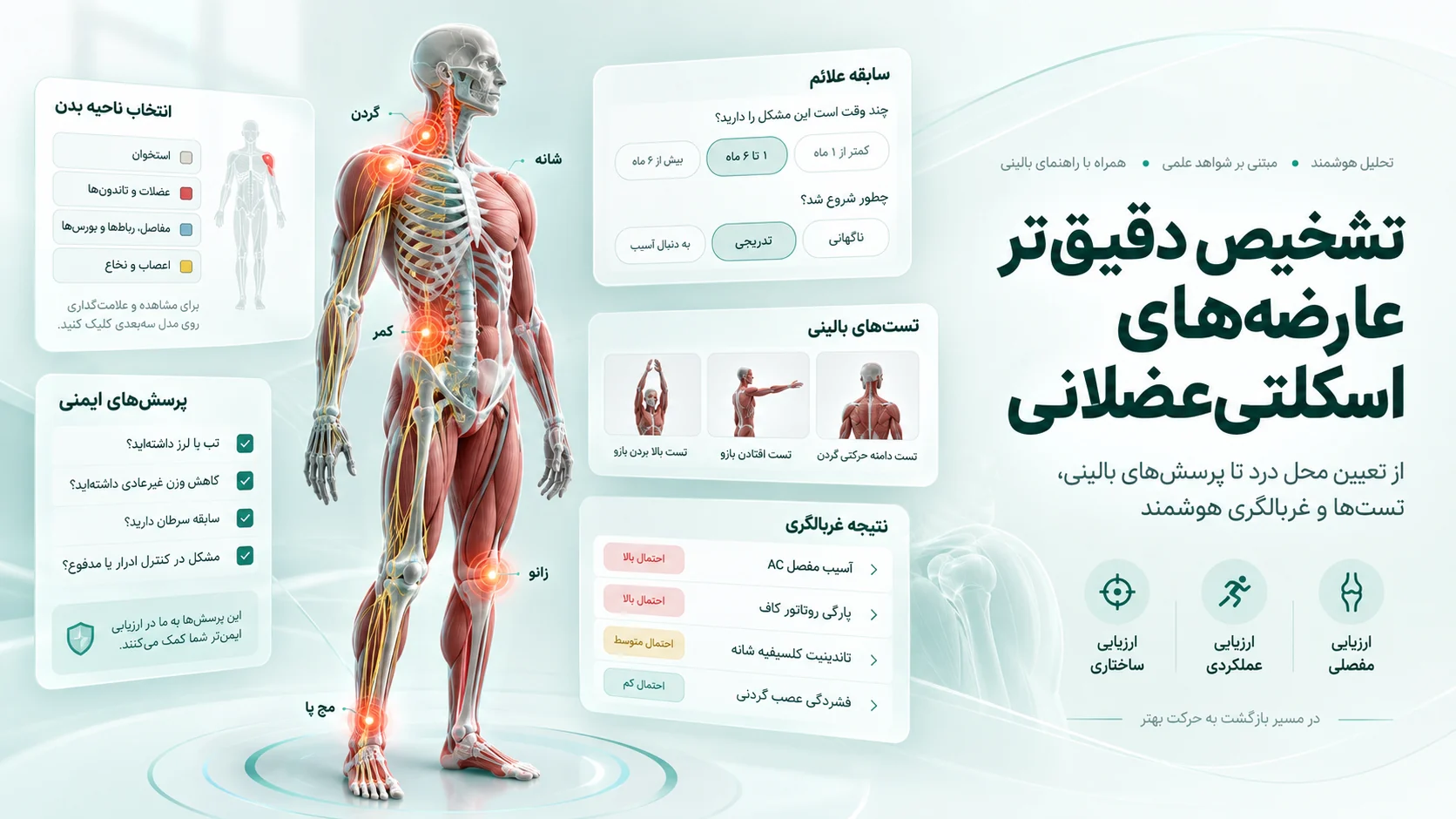
- Mark the painful regionYou select the region that hurts on a 3D skeleton. The model is anatomically real and you can rotate it to find the exact spot.
- The matching test setEach region calls up its own test protocol — not a generic questionnaire.
- Guided testingEvery test is shown with an image: how to stand, what to do, and what you should feel. Your answer (painful / not painful / unsure) is recorded.
- ScoringThe pattern of your answers is compared against the known pattern for each condition, and findings are listed in order of likelihood.
- Deciding the next stepThe report states plainly which findings can be followed with corrective exercise and which need a physician's examination or imaging.
Regions and conditions covered
Spine
Lumbar and cervical disc herniation, excessive lumbar curve, rounded upper back, lateral spinal deviation, and the syndromes that follow from imbalance in the trunk muscles.
Shoulder and upper limb
Subacromial impingement, rotator cuff problems, rounded shoulders and restricted scapular range.
Hip and knee
Knee osteoarthritis, meniscal and cruciate ligament injury, patellofemoral pain, pelvic drop and rotation, hip osteoarthritis and short hip flexors.
Ankle and lower leg
Achilles tendinopathy, ankle instability, flat foot and its effect on the chain above it.
When the pain is not where the problem is
One reason a region-only assessment is not enough is that the body is a connected chain. Restricted ankle range makes the knee take more load in sitting and standing. Weak hip abductors let the knee fall inward on landing. Tight hip flexors tilt the pelvis forward and deepen the lumbar curve.
In all of those examples, where it hurts and where the correction belongs are not the same place. That is why musculoskeletal screening is usually read alongside posture analysis and, where relevant, gait analysis: the clinical tests narrow down where the problem is, and the posture and gait measurements show what has been holding it there.
Common patterns and how they usually present
The table below shows patterns that come up often in screening. These are typical presentations, not diagnostic criteria:
| Region | Common pattern | Typical presentation |
|---|---|---|
| Neck | Forward head with lower neck pain | Worse by the end of a work day, better lying down |
| Shoulder | Subacromial impingement | Pain raising the arm between roughly 60 and 120 degrees |
| Lower back | Disc involvement | Worse bending forward and sitting for long periods, may radiate down the leg |
| Lower back | Facet joint involvement | Worse leaning back and rotating, stays local |
| Hip | Weak abductors | Pelvic drop when standing on one leg |
| Knee | Patellofemoral pain | Worse descending stairs and sitting with the knee bent |
| Ankle | Achilles tendinopathy | Morning stiffness, pain at the start of activity |
What a clinical test says, and what it does not
Every clinical test has two properties: sensitivity (how well it catches true cases) and specificity (how well it rules out the ones that are not). No test is excellent at both. That is why a single positive test means little, while three positive tests all pointing to one mechanism mean something.
This assessment is screening. Its output is a ranked likelihood, not a diagnosis. A definitive diagnosis needs an in-person examination and, in many cases, imaging.
Signs that mean see a doctor now
If you have any of these, stop the online assessment and see a physician:
- Progressive numbness or weakness in an arm or leg
- Loss of bladder or bowel control together with back pain
- Severe night pain that rest does not relieve
- Pain after a fall or impact, with swelling or inability to bear weight
- Fever together with joint pain
- Unexplained weight loss together with musculoskeletal pain
What happens afterwards
The report, with its findings and suggested programme, is yours. If the findings sit within the scope of corrective exercise, a phased programme with an image for every exercise is provided. If they do not, the report says plainly that the right path is a physician — and you can take that same report with you, so your doctor knows what has already been checked.
Frequently asked questions
Will this tell me whether I have a disc herniation?
No. Clinical tests raise or lower the likelihood of disc involvement, but confirming or excluding it needs a physician's examination and, in many cases, an MRI. The report states this explicitly.
Is it risky to perform the tests myself?
The tests selected are all active tests you perform yourself and are low-risk. If a test causes severe pain you should stop — that instruction appears with every test. Tests that require an assessor's hands are done during an in-person session.
How long does it take?
Depending on the region, usually ten to twenty minutes.
Do I have to travel to Quchan for this assessment?
Not for the online screening — the tests you perform yourself can be done from anywhere. The tests that need an assessor's hands, and the full examination, are done at the clinic in Quchan, Razavi Khorasan.
Can I take the report to my doctor?
Yes, the report is printable and designed for exactly that. Findings are written with the standard names of the tests so a clinician can read them.