
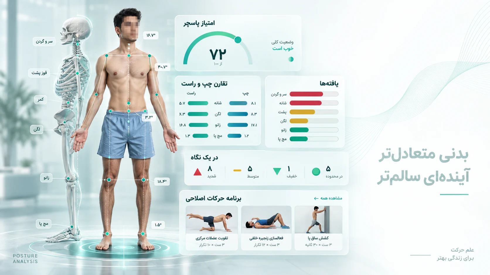
What posture is, and why looking is not enough
Posture is the position your body settles into when standing at rest. It is the net result of muscle length, tissue stiffness, bone shape and the way your nervous system organises all of it. The problem with assessing it by eye is that the human eye misses small differences and exaggerates large ones. Two assessors describe the same person differently — and, more importantly, when you want to know three months later whether the exercise programme did anything, you have no baseline number to compare against.
The fix is to turn observation into measurement. Once your forward head angle has a number today, that same number can be taken again in three months and compared. The value of posture analysis is that it repeats, not that it puts a label on you.
Two paths: online screening and the in-person session
Posture assessment here runs on two paths, and they are not the same thing:
- Online screening. You take three photographs with your own phone and view a free summary without an account. The full report requires payment and active account access. This path works from anywhere in Iran, and what it produces is a measurement, not a diagnosis.
- The in-person session in Quchan. Examination, functional testing, pain history, and a corrective exercise programme written for you. People travel in from Quchan and the towns around it — Faruj, Dargaz and Chenaran — and from Mashhad.
The usual order is screening first, then bringing that report to the in-person session. Neither path replaces a physician's examination or medical imaging.
What gets measured
The assessment runs on three standard views, and each view carries its own set of measures. The table below lists the main ones alongside their method-specific interpretation notes. This table does not define universal diagnostic thresholds: interpretation depends on the capture method, landmarks and individual context. A photograph alone cannot establish the cause of pain.
| Measure | View | Method and interpretation | What it indicates |
|---|---|---|---|
| Craniovertebral angle (CVA) | Lateral | Depends on angle definition and capture method | Forward head posture |
| Thoracic surface curvature | Lateral | Method and landmark dependent | Not radiographic kyphosis |
| Lumbar surface curvature | Lateral | Method and landmark dependent | Not radiographic lordosis |
| Surface trunk and back alignment | Posterior | No diagnostic Cobb threshold | Surface asymmetry, not a Cobb measurement |
| Shoulder height difference | Anterior / posterior | Metric distances require a valid scale | Shoulder girdle asymmetry |
| Pelvic landmark height difference | Posterior | Metric distances require a valid scale | Pelvic drop or rotation |
| Q angle | Anterior | Depends on landmarks, limb position and method | Knee alignment, valgus |
| Rearfoot pronation angle | Posterior | Depends on capture view and heel-axis definition | Inward roll of the ankle |
The report includes measures supported by the available input. Missing views or usable landmarks limit what can be assessed. Read the photogrammetry evidence and limitations and the difference between screening and diagnosis.
The three views
Anterior — from the front
Horizontal alignment of shoulders, pelvis and knees, internal or external rotation of the legs, and the Q angle come from this view. Knock knees (valgus) and bow legs (varus) are seen here.
Lateral — from the side
The most informative view for the spine. A plumb line is drawn from the ear to the lateral malleolus and the deviation of head, shoulder, pelvis and knee from that line is measured. This view describes external back contours and head and shoulder alignment; these measurements are not vertebral angles on radiographs.
Posterior — from behind
Symmetry between the two halves of the body: lateral spinal deviation, scapular height difference, pelvic rotation and rearfoot position.
The calibration sheet — optional, but it takes scale out of guesswork
Angles need no scale: the ratio between two lines is the same whatever size the photograph is. Distances are different. To say "18 millimetres", something has to establish how many millimetres a pixel is, and with no object of known size in frame that comes from the height you typed in — an assumption rather than a measurement.
The calibration sheet carries two black squares of exactly known size. Lay it on the floor and keep it in frame, and three things are read off the photograph instead of being assumed: true scale from the size of the squares, the floor line from the line between them, and the room's true vertical — which matters, because the vertical edge of a photograph is only plumb if the camera was perfectly level, and it usually was not.
The printable calibration sheet (A2) — print it on A2 at 100% scaling ("Actual size"), then check with a ruler that the black square measures 120 mm. If the printer shrank it, every distance is wrong by the same proportion and nothing in the report would reveal it; the sheet carries that warning, and how to check it, printed on itself.
Some measurements remain possible without the sheet, but the scale method and units must be clear. Do not interpret pixel distances as centimetres without a usable scale. Review the capture and scaling checklist before starting.
How the assessment runs
- Standardised photographsThree views, with fixed camera distance and height, suitable clothing and a plain background. The shooting conditions are explained on the page — ignoring them invalidates the measurement.
- Landmark detectionA computer-vision model locates the anatomical reference points on the image.
- Angles and distancesThe tool measures lines between image landmarks and distances from image references. Two-dimensional photograph measurements are not equivalent to in-person goniometry or vertebral angles on radiographs.
- Clinical reviewA number on its own is not a diagnosis. The findings are read in person alongside examination, pain history and functional testing.
- Corrective exercise programmeExercises are chosen from the measures that fell outside their reference range, with a set interval before the next measurement.
What the report contains
- The three view photographs with landmarks and measurement lines drawn on them
- A numeric table of every measure beside its reference range
- The findings that fell outside range, ordered by importance
- An exercise programme matched to those findings
- The assessment date, so the next measurement is comparable
The report is printable and you can take it to your own physician or physiotherapist. Before you start, see the sample client and practitioner reports, built from synthetic data.
Who benefits from this
- People who sit at a desk for hours and have neck, shoulder or back pain.
- Adolescents whose parents suspect a spinal curve or uneven shoulders — an initial screen, before deciding about radiography.
- Athletes who keep injuring the same side and want to know whether a structural asymmetry is involved.
- Anyone already following an exercise programme who wants to know whether anything has actually changed after a few months.
If you are a coach or practitioner assessing your own clients, see the practitioner workflow.
There is no single "correct" posture
The common assumption is that one ideal posture exists and every deviation from it is a problem. The evidence does not support that: normal variation between people is wide, and many people whose posture sits far from average have no pain at all. The relationship between posture and pain is not simple or one-to-one.
So why measure? For three reasons. First, to have a baseline to measure change against. Second, to find asymmetries — which matter more than deviation from average, because a body is usually symmetrical with itself. Third, to see whether the mechanical finding lines up with the complaint; when the painful site and the measured finding agree, the exercise programme has a clear target.
For that reason, no single number in the report is called "bad" on its own. Numbers are read next to your complaint.
Where this assessment stops
Posture analysis is a screening and measurement tool, not a substitute for medical diagnosis. Measuring from a photograph captures surface alignment and does not replace radiography, MRI or a physician's examination. Surface curvature on a photograph is not a radiographic Cobb angle. The Scoliosis Research Society describes Cobb measurement on spinal X-rays.
If you have night pain, numbness, muscle weakness or a sudden change in posture, a physician should examine you before any exercise programme begins.
Free summary or full report?
| Output | Access |
|---|---|
| Online screening summary | Free; start without an account |
| Full posture report | 790,000 toman; account and access activated after payment |
| Full musculoskeletal screening report | 500,000 toman; separate from posture analysis |
| Individual practitioner interpretation and programme | Requires practitioner assessment and the relevant service fee; distinct from the automated summary |
A single report unlocks that assessment; practitioner plans have separate terms. See the online services shop. If payment is unavailable in your account, contact the centre. Creating an account alone does not unlock the full report. Fees and arrangements · Sign in
Frequently asked questions
Do I have to come in person for posture analysis?
Not to get the numeric report. You run the initial screening yourself, with three photographs, inside the browser on your own device, from anywhere. Clinical interpretation, examination and a personalised exercise programme require an in-person session at the clinic in Quchan.
Where is the clinic, and where do people travel from?
The clinic is in Quchan, Razavi Khorasan, Iran: Unit 3, Parseh Complex, Nikooei St. Most people come from Quchan and the towns around it — Faruj, Dargaz and Chenaran — and from Mashhad. The online screening has no such limit.
How long does the online screening take?
Photographing and measuring are usually done in one sitting. Most of the time goes on meeting the shooting conditions, because an incorrect camera distance or height invalidates the measurement.
Does this replace an X-ray?
No. Measuring from a photograph captures surface alignment. Measuring spinal curvature precisely, or assessing bone structure, requires radiography, and that is a physician's call.
How often should I repeat the assessment?
Retesting depends on the assessment purpose, symptoms and the practitioner’s follow-up plan; one fixed interval does not fit everyone. Keep capture conditions and methods consistent. A numerical change alone does not establish clinical improvement.
Can adolescents be assessed?
Yes, and screening spinal symmetry matters more during growth years. But if a significant deviation is found, the next step is a physician, not starting exercises on your own.