
What it means
A sideways curve that disappears on sitting or when the leg length is equalised.
Where it comes from
The spine's response to a tilted pelvis or muscle spasm, not a structural change.
How it is measured
It is measured on the back view, in the coronal plane. The defining measurement is Sideways curve of the spine (Cobb angle), and it is reported as a finding from above 10°. The number is computed from landmarks on that photograph — not estimated by eye, and not compared against a stock example.
What the numbers mean
| Measurement | Grade |
|---|---|
| up to 10° | No scoliosis |
| 10–25° | Mild |
| 25–40° | Moderate |
| 40° and above | Severe |
Source of the bands: Surface curve magnitude, SRS bands; the functional/structural split is from the pelvic reading, not the curve
What is usually seen alongside it
- One hip higher (Pelvic obliquity) — above 4°
- One shoulder higher (Acromial tilt) — above 2°
None of these is a diagnosis on its own. Together they corroborate the finding; apart, they do not refute it — a measurement nobody took counts neither for nor against.
What confirms it
If the curve disappears on sitting or once the leg lengths are equalised, it is functional. A tape measurement from the ASIS to the medial malleolus settles it.
Functional scoliosis — a curve that is not structural
A functional scoliosis is a lateral curve that does not come from the spine itself but is a response to something else — and that is the difference that matters: resolve the cause and the curve resolves.
The cause most often looked for
A leg-length difference. Knutson [2], using accurate radiographic methods, found anatomic inequality in 90% of people, with a mean of 5.2 mm — so the mere presence of a difference explains nothing. What matters is its size, and the evidence suggests it does not become clinically significant for most people until around 20 mm.
And the distinction that sets the order of work
Part II of the same review [1] holds that the functional "short leg" — what is seen lying down, unloaded — is a different phenomenon from anatomic inequality, and may come from suprapelvic muscle hypertonicity. The two interact in standing and not when lying.
⚠️ Its practical recommendation: functional asymmetry should be resolved before any treatment of an anatomic inequality. Done the other way round, something may get compensated for that was never bone.
How it is separated from structural
By examination, not from a photograph. A functional curve usually reduces or disappears with a change of position — bending forward, sitting, or compensating the leg-length difference; a structural one does not, and carries a rotational component. The structural scoliosis page has the figures decisions rest on.
⚠️ During growth, telling the two apart is a doctor's work and may need imaging. A photographic screen only says whether asymmetry is present.
References
- Knutson GA. Anatomic and functional leg-length inequality: a review and recommendation for clinical decision-making. Part II, the functional or unloaded leg-length asymmetry. Chiropr Osteopat. 2005;13:12. PMID 16080787 · DOI 10.1186/1746-1340-13-12
- Knutson GA. Anatomic and functional leg-length inequality: a review and recommendation for clinical decision-making. Part I, anatomic leg-length inequality: prevalence, magnitude, effects and clinical significance. Chiropr Osteopat. 2005;13:11. PMID 16026625 · DOI 10.1186/1746-1340-13-11
The evidence on this
Study summaries that measured this finding — with their numbers and their limits.
- Scoliosis-specific exercise and the Cobb angle — Systematic review and meta-analysis, 18 randomised trials
The exercise programme
A lateral spinal curve is a three-dimensional problem and its exercise must be asymmetric and supervised. Symmetrical training does not help here.
⚠️ A curve beyond about 20° needs an in-person assessment and an X-ray. A scoliosis programme must be written specifically for the side of convexity and the level of the apex.
The stages advance on a standard, not on the calendar:
- Stage 1 — release and wake up — weeks 1–4: Quadratus lumborum stretch
Move on: When the movement is done without pain and without compensating, and the stretch is no longer what limits it. - Stage 2 — control — weeks 3–8: Side plank, Quadratus lumborum stretch
Move on: When the target muscle is felt through the whole range and the last set looks like the first. - Stage 3 — load and integration — weeks 6–16: Lateral elongation for scoliosis, Diaphragmatic breathing, Side plank
Move on: When the corrected pattern holds in everyday movement and under fatigue.
Each session 3–5 times a week, for at least 12 weeks. (from a study)
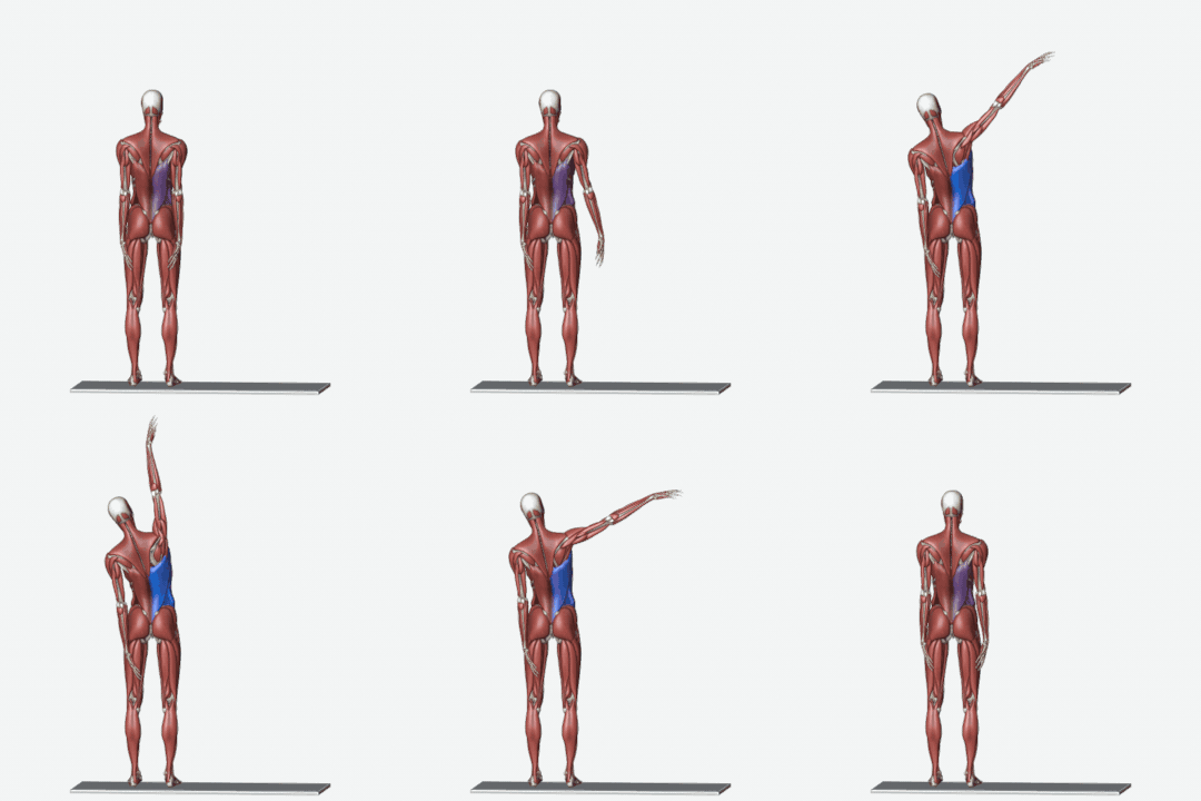
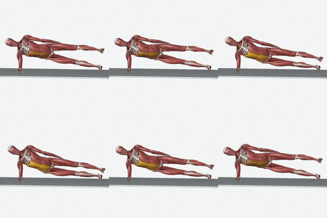
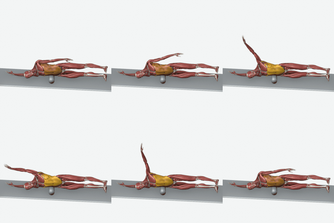
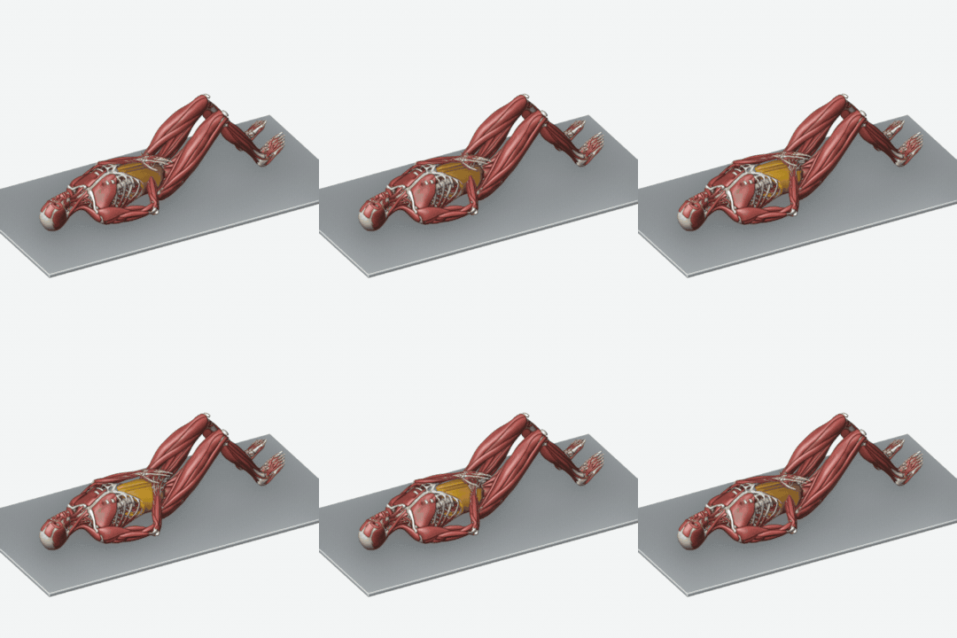
This example is for a moderate grade; each stage runs shorter or longer with the measured grade, and a person's own programme is written after assessment, around their other conditions and limits. If a movement causes pain, stop it and contact the clinic. Muscle colours in the animations: orange working, blue stretching, amber holding still.
Every number on this page is measured on the skin surface in a photograph. That is not the same as a measurement taken on a radiograph: it correlates with one and does not replace it. Wherever the decision depends on imaging, this page and the report itself say so.
What happens at the clinic
The assessment starts with three standing photographs — front, side and back. Every angle is measured on those photographs, the report prints the number and the grade for each, and the exercise programme is built from those numbers rather than from a template. Posture analysis explains the whole path, and the online assessment can be started from your own photograph now.
Frequently asked questions
At what point is functional scoliosis reported?
When sideways curve of the spine (cobb angle) is above 10°, it is reported as a finding. That is where the "Mild" band begins. The bands come from: Surface curve magnitude, SRS bands; the functional/structural split is from the pelvic reading, not the curve
Can functional scoliosis be assessed from a photograph?
A photograph gives a number, which most assessments do not have. But it is a surface measurement and it does not replace imaging. If the curve disappears on sitting or once the leg lengths are equalised, it is functional. A tape measurement from the ASIS to the medial malleolus settles it.
What is the most severe grade?
The worst band in this table is "Severe". The grade is printed beside the measurement itself in the report, so it is always clear which number produced the word.